
Trace the journey of a zygote from the isthmus of the fallopian tube up to its implantation in the uterus of a human female. Highlight the changes the zygote undergoes during the course of its journey up to implantation.
Correct Answer :
Mitotic division of the zygote (in isthmus) is called cleavage, the zygote moves towards the uterus, cleavage results in formation of (2,4,8,16) daughter cells called blastomeres, embryo with 8 to 16 blastomeres is called morula which continues to divide and transforms into blastocyst as it moves further into the uterus, blastomeres in the blastocyst are arranged into an outer layer called trophoblast, trophoblast layer gets embedded to the endometrium and this is called implantation.
//
Solution :
Correct Answer/Option:
Mitotic division of the zygote (in the isthmus) is called cleavage; the zygote moves towards the uterus. Cleavage results in the formation of 2, 4, 8, and 16 daughter cells called blastomeres. The embryo with 8 to 16 blastomeres is called a morula, which continues to divide and transforms into a blastocyst as it moves further into the uterus. The blastomeres in the blastocyst are arranged into an outer layer called the trophoblast. The trophoblast layer gets embedded in the endometrium, and this process is called implantation.
Step-by-Step Explanation:
1. Fertilization and Zygote Formation:
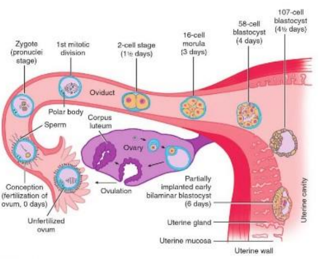
The process begins at day 0 in the fallopian tube (oviduct). As labeled in the provided diagram, the fusion of a sperm with the "Unfertilized ovum" leads to "Conception (fertilization of ovum, 0 days)". This event forms a single-celled diploid "Zygote (pronuclei stage)".
2. Cleavage and Blastomere Formation:
As the zygote begins to travel through the fallopian tube towards the uterus, it undergoes rapid mitotic divisions known as cleavage. The first division is depicted in the diagram as the "1st mitotic division" (accompanied by a "Polar body"), which quickly yields the "2-cell stage" at approximately 11/2 days. Subsequent divisions produce 4, 8, and then 16 smaller daughter cells called blastomeres.
3. Transition to the Morula Stage:
By day 3, the cluster of dividing blastomeres forms a solid ball of cells resembling a mulberry. The diagram labels this stage as the "16-cell morula".
4. Blastocyst Development:
As the morula moves further through the "Oviduct" towards the "Uterine cavity", it undergoes differentiation and forms a fluid-filled cavity. The embryo is now called a blastocyst. The diagram marks this transition through the "58-cell blastocyst (4 days)" and the "107-cell blastocyst (41/2 days)" stages. During this time, the blastomeres organize into two distinct regions:
• An outer cellular layer called the trophoblast.
• An inner group of cells known as the inner cell mass (which will eventually develop into the embryo itself).
5. Implantation:
Upon reaching the uterus, the blastocyst prepares to attach. As shown in the diagram at day 6, the outer trophoblast layer of the "Partially implanted early bilaminar blastocyst (6 days)" begins to embed into the endometrium (labeled as "Uterine mucosa") near the "Uterine gland" along the "Uterine wall". This successful embedding is called implantation, which establishes the connection necessary for maternal nutrient exchange and further development.
Access expert-curated educational resources and study materials—completely free.
Create, conduct, and manage professional online assessments with Mindyard. Perfect for teachers and institutes.
Copyright © 2026 Mindyard. All Rights Reserved.
 Set - 3-1782888548748.webp)